hospitals in Florence and Rome and in Paris (France)
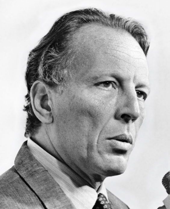

Filho da educadora em saúde Anna Bove e do dentista Alfredo Pinotti, José Aristodemo Pinotti, ou Ari como era chamado pela família, nasceu em um ambiente propício para seguir sua vocação na área médica. Todos os seus avós vieram da Itália. Seus avós paternos eram de Veneto, enquanto seus avós maternos da região de Amalfi.
Ele visitou a Itália pela primeira vez em 1961, e a partir de então criou profundos laços profissionais e de amizade lá. Tanto que sua formação médica se aprimorou no período em que passou internados em hospitais de Florença e Roma e em Paris (França), logo após o término do curso na Faculdade de Medicina da Universidade de São Paulo. Por fim, ficou em Milão, no então Istituto dei Tumori, onde conheceu um jovem cirurgião de mama chamado Umberto Veronesi, com quem manteve uma profunda amizade e parceria profissional que rendeu muitos frutos para a mastologia brasileira.
Nessa época, já almejava a carreira universitária. Convidado pelo Prof. Bussâmara Neme, seu professor de Medicina e também professor da Faculdade de Ciências Médicas da Universidade Estadual de Campinas (Unicamp), para criar o Departamento de Ginecologia e Obstetrícia daquela faculdade, percebeu a grande oportunidade de iniciar seus objetivo principal: tornar-se professor universitário de medicina.
Nas décadas de 1970 e 1980 deu importantes contribuições para garantir melhores possibilidades de cura e qualidade de vida às mulheres com câncer de mama, sendo pioneiro em pesquisas sobre quimioterapia adjuvante com seu companheiro de profissão e de vida Prof. Dr. Luíz Carlos Teixeira. Na década de 1980 publicou com os Profs. Henrique Benedito Brenelli, Prof. Maurício Knobel e Prof. Ricardo Baroudi um importante trabalho sobre reconstrução mamária imediata pós-mastectomia que foi transformador em nossa conduta. Foi também quando iniciou os protocolos de cirurgia conservadora com avaliação citohistológica das margens cirúrgicas, que atualmente nos permitem utilizar as técnicas de mamoplastia.
Toda a sua carreira acadêmica foi realizada na Unicamp, onde prestou os exames que o levaram ao cargo de professor titular. Também por meio de concursos, alcançou o mesmo cargo na Faculdade de Medicina da USP. Foi Reitor da Faculdade de Medicina e Reitor da UNICAMP quando construiu e fundou o Centro de Atenção Integral à Saúde da Mulher (CAISM) que hoje leva seu nome (Prof. José Aristodemo Pinotti Hospital da Mulher) e que é reconhecido em todo o Brasil.
Sua relevante contribuição científica está traduzida em 82 livros e 1.860 artigos científicos publicados em renomadas revistas internacionais, muitas delas premiadas.
Na década de 1960 reuniu uma equipe para montar sua primeira clínica médica privada voltada para o atendimento integral às condições de saúde da mulher.
Como gestor público, ampliou o conceito de universalização e integração da saúde em sua extensa obra: foi reitor da Unicamp (1982-1986), secretário estadual de Educação e Saúde (1986-1991), deputado federal por três legislaturas, membro do Academia Nacional de Medicina (cadeira 22), secretário municipal de Educação de São Paulo (2005-2006), único brasileiro eleito presidente da Federação Internacional de Ginecologia e Obstetrícia (1986-1992), e o primeiro a receber o título de Doutor Honoris Causa pela Universidade de Bolonha, a mais antiga do mundo, em reconhecimento à importância de seu trabalho em prol das mulheres.
Em 1966 casou-se com a artista plástica e professora do Instituto de Artes da Unicamp, Suely Pinotti, com quem construiu sua família que sempre o apoiou e colaborou em todos os seus projetos. Teve três filhos: Marianne (médica), André (arquiteto) e Mirella que faleceu aos 19 anos. Teve cinco netos: Anna, Gaia, Ari, Enrica e Mirella.
Admirador e entusiasta da literatura, da música e do teatro, publicou dois livros de poemas. Conviveu com artistas do mundo inteiro, como a poetisa e escritora Hilda Hilst e José Antônio de Almeida Prado, grande compositor da música erudita contemporânea, entre outros.
Livre pensador e ávido leitor, com imensa capacidade de síntese e generosa difusão de ideias e utopias, conciliou a sua vocação científica com o seu carácter intensamente humanista.
O prêmio José Aristodemo Pinotti é concedido ao melhor artigo apresentado durante o BBCS. Inicialmente, tratamos das passagens aéreas, hospedagem e inscrição da autora no San Antonio Breast Cancer Symposium, no Texas (EUA). Porém, com a pandemia e o evento online, a edição de 2021 entregou o prêmio em dinheiro. A edição de 2022 seguirá o mesmo formato.